Medical Image Segmentation
751 papers with code • 44 benchmarks • 43 datasets
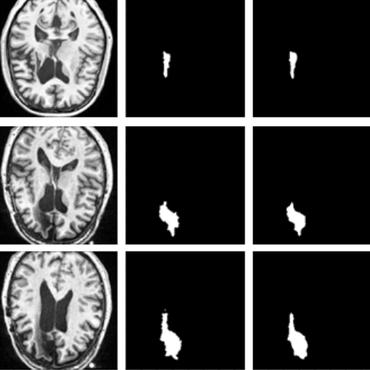
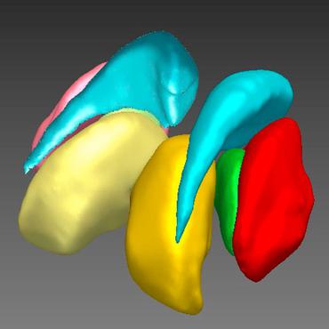
Medical Image Segmentation is a computer vision task that involves dividing an medical image into multiple segments, where each segment represents a different object or structure of interest in the image. The goal of medical image segmentation is to provide a precise and accurate representation of the objects of interest within the image, typically for the purpose of diagnosis, treatment planning, and quantitative analysis.
( Image credit: IVD-Net )
Libraries
Use these libraries to find Medical Image Segmentation models and implementationsDatasets
Subtasks
-
 Lesion Segmentation
-
 Brain Tumor Segmentation
-
 Cell Segmentation
-
 Skin Lesion Segmentation
-
 Skin Lesion Segmentation
-
 Brain Segmentation
-
 Retinal Vessel Segmentation
-
 Semi-supervised Medical Image Segmentation
-
 MRI segmentation
-
 Cardiac Segmentation
-
 3D Medical Imaging Segmentation
-
 Liver Segmentation
-
 Volumetric Medical Image Segmentation
-
 Brain Image Segmentation
-
 Pancreas Segmentation
-
 Iris Segmentation
-
 Video Polyp Segmentation
-
 Lung Nodule Segmentation
-
 Nuclear Segmentation
-
 COVID-19 Image Segmentation
-
 Skin Cancer Segmentation
-
 Electron Microscopy Image Segmentation
-
 Ischemic Stroke Lesion Segmentation
-
 Brain Lesion Segmentation From Mri
-
 Placenta Segmentation
-
 Infant Brain Mri Segmentation
-
 Automatic Liver And Tumor Segmentation
-
 Acute Stroke Lesion Segmentation
-
 Cerebrovascular Network Segmentation
-
 Automated Pancreas Segmentation
-
 Semantic Segmentation Of Orthoimagery
-
 Pulmorary Vessel Segmentation
-
 Brain Ventricle Localization And Segmentation In 3D Ultrasound Images
Most implemented papers
U-Net: Convolutional Networks for Biomedical Image Segmentation
There is large consent that successful training of deep networks requires many thousand annotated training samples.
Densely Connected Convolutional Networks
Recent work has shown that convolutional networks can be substantially deeper, more accurate, and efficient to train if they contain shorter connections between layers close to the input and those close to the output.
An Image is Worth 16x16 Words: Transformers for Image Recognition at Scale
While the Transformer architecture has become the de-facto standard for natural language processing tasks, its applications to computer vision remain limited.
SegNet: A Deep Convolutional Encoder-Decoder Architecture for Image Segmentation
We show that SegNet provides good performance with competitive inference time and more efficient inference memory-wise as compared to other architectures.
Attention U-Net: Learning Where to Look for the Pancreas
We propose a novel attention gate (AG) model for medical imaging that automatically learns to focus on target structures of varying shapes and sizes.
UNet++: A Nested U-Net Architecture for Medical Image Segmentation
Implementation of different kinds of Unet Models for Image Segmentation - Unet , RCNN-Unet, Attention Unet, RCNN-Attention Unet, Nested Unet
V-Net: Fully Convolutional Neural Networks for Volumetric Medical Image Segmentation
Convolutional Neural Networks (CNNs) have been recently employed to solve problems from both the computer vision and medical image analysis fields.
TransUNet: Transformers Make Strong Encoders for Medical Image Segmentation
Medical image segmentation is an essential prerequisite for developing healthcare systems, especially for disease diagnosis and treatment planning.
Brain Tumor Segmentation with Deep Neural Networks
Finally, we explore a cascade architecture in which the output of a basic CNN is treated as an additional source of information for a subsequent CNN.
Recurrent Residual Convolutional Neural Network based on U-Net (R2U-Net) for Medical Image Segmentation
In this paper, we propose a Recurrent Convolutional Neural Network (RCNN) based on U-Net as well as a Recurrent Residual Convolutional Neural Network (RRCNN) based on U-Net models, which are named RU-Net and R2U-Net respectively.


 DRIVE
DRIVE
 Kvasir-SEG
 Kvasir
 GlaS
GlaS
 Medical Segmentation Decathlon
 PROMISE12
 AMOS
AMOS
 CHASE_DB1
 ACDC
ACDC
 CVC-ClinicDB


